Retinal Diagnostic Station
Retinal Diagnostic Station
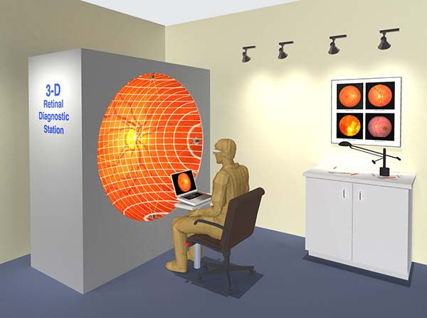
There has not been a satisfactory way to view the images to preserve the angular relationships until the HinesLab Retinal Diagnostic Station which displays stereoscopic images to provide the point of view as if the ophthalmologist were perched in the patient’s pupil, looking around at the very realistic interior of the eye, all in 3D.
Several eye diseases can be detected with early examination of the retina. Ophthalmologists use a fundus camera to photograph the retinas with a field view up to 140°. When these pictures are taken stereoscopically much more information is conveyed about the spatial relationships of the veins, floaters and any detachment of the retina. The pictures below were taken with the Topcon NW400 retinal camera which can automate the taking of stereoscopic pairs.
The 3-D picture is projected into the concave screen which simulates the interior of the patient’s eye. Ophthalmologists wear 3-D glasses and see the features in the eye in a 1:1 angular relationship, making diagnosis more intuitive.
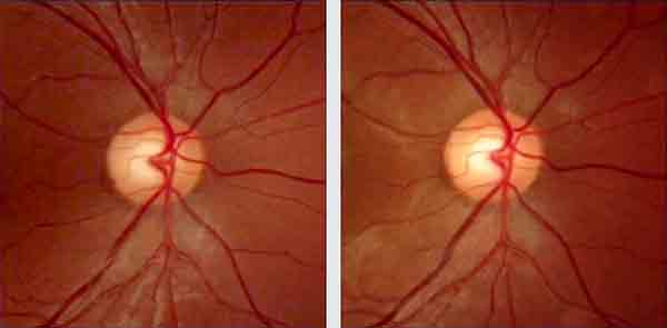 Left Right
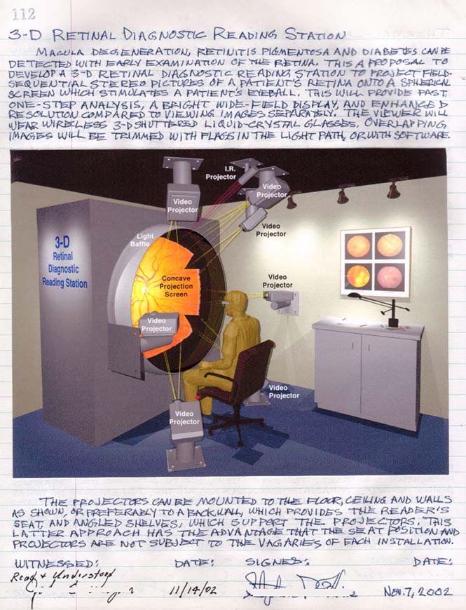
Steve Hines’ entry, Notebook #1, p. 112:
Comments:
- “I am very impressed with the wrap-a-round system you have developed for viewing retinal images.” Nov. 15, 2018, Dr. Patricia Bath, Ophthalmologist
HinesLab is actively seeking licensees to commercialize this technology. Please contact Steve Hines at: