Moiré Microscope
Moiré Microscope
 |
|
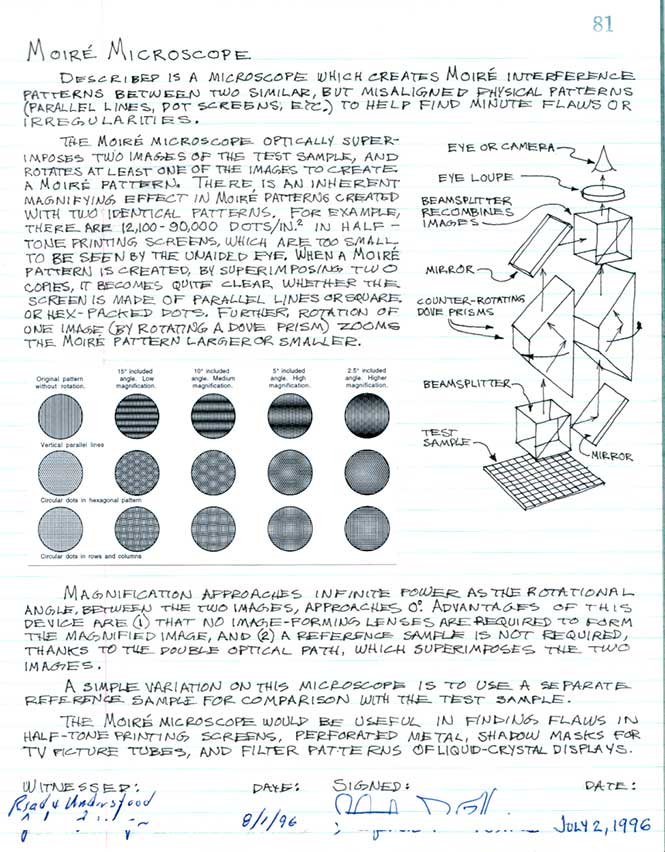
The moiré microscope helps identify the geometric structure of material samples. For example, fly’s eyes, graphene, and fiber-optic image bundles have 6-axis or hexagonal patterns. Calcite (salt), and pyrite have a 90° square-pack arrangement. The microscope optically super-imposes the image of the sample onto a reference pattern (the yellow ring) which is rotated to create a moiré* pattern. It becomes quite clear whether the sample has a hexagonal, square-packed or parallel-line structure. *Moiré, French for “watery”.
.png)
|
Moiré imagery can be used to compare a sample to itself, using dove prisms and mirrors create a second image that can be superimposed and rotated over the original sample for comparison. For example, half-tone printing screens have from 12,000 – 90,000 hex-packed dots in each square inch. When viewed with the moiré microscope, the hex pattern is immediately apparent. Magnification reaches infinite optical power (although inherently out of focus) as the two patterns are aligned, not possible in a conventional microscope. |
 |
_-_schematic.svg)
The value of the moiré microscope is as a field instrument to quickly categorize the geometric structure of samples.
Hines’ lab notebook #1, p. 81:
Reference: A moiré microscope for finite deformation micro-mechanical studies
Experimental Mechanics volume 40, pages 351–360 (2000), $39.95
HinesLab is actively seeking licensees to commercialize this technology. This is not a product for sale. To discuss licensing, please contact Steve Hines at: